3D image processing program
Quantification and visualization of changes in alveolar bone resorption over time
Overview
Periodontitis, which causes resorption of alveolar bone, affects most adults and causes tooth loss as it progresses. In recent years, dental Cone Beam CT has become popular, enabling three-dimensional confirmation of the morphology of alveolar bone. However, in most cases, only visual qualitative morphological evaluation was performed, and there was no method to detect minute morphological changes over time or to quantitatively analyze the amount of changes. The present invention makes it possible to visualize and quantify changes in the surrounding bone morphology by performing precise alignment using morphological information of only the root portion of any tooth.
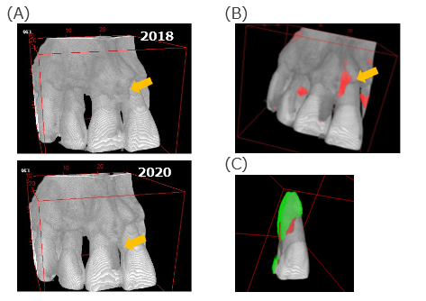
FIG. A shows alveolar bone CT images of the same patient taken at different times, but it is difficult to confirm at a glance the exact area where bone was absorbed (yellow arrow) and the amount of bone resorption over a period of two years. By performing a semi-automatic analysis of about 10 seconds using the program of this invention, it is possible to display the resorbed bone in red (FIG. B) and calculate the amount of bone resorption by volume. As shown in FIG. C, it is also possible to color the root surface of the tooth to discriminate the areas where bone is still covered (green) and where bone is lost due to resorption (red).
Features・Outstandings

Product Application
・Application to dental Cone Beam CT equipment
・Application to CT or MRI equipment for evaluating bone resorption around artificial joints
IP Data
IP No. : PCT/JP2022/035892
Inventor : YAMAGUCHI Satoshi, NAKAMURA Megumi
keyword : bone resorption、quantitive analysis and visualization、 3D imaging
